
The 64-channel system
The 64-channel system is a research device that records muscle activity from 64 points at the same time. It is being designed for clinicians and researchers who study the pelvic floor, trunk and limbs, including in stroke rehabilitation.
In development. Not for sale. Not cleared or approved by the FDA or any other regulator.
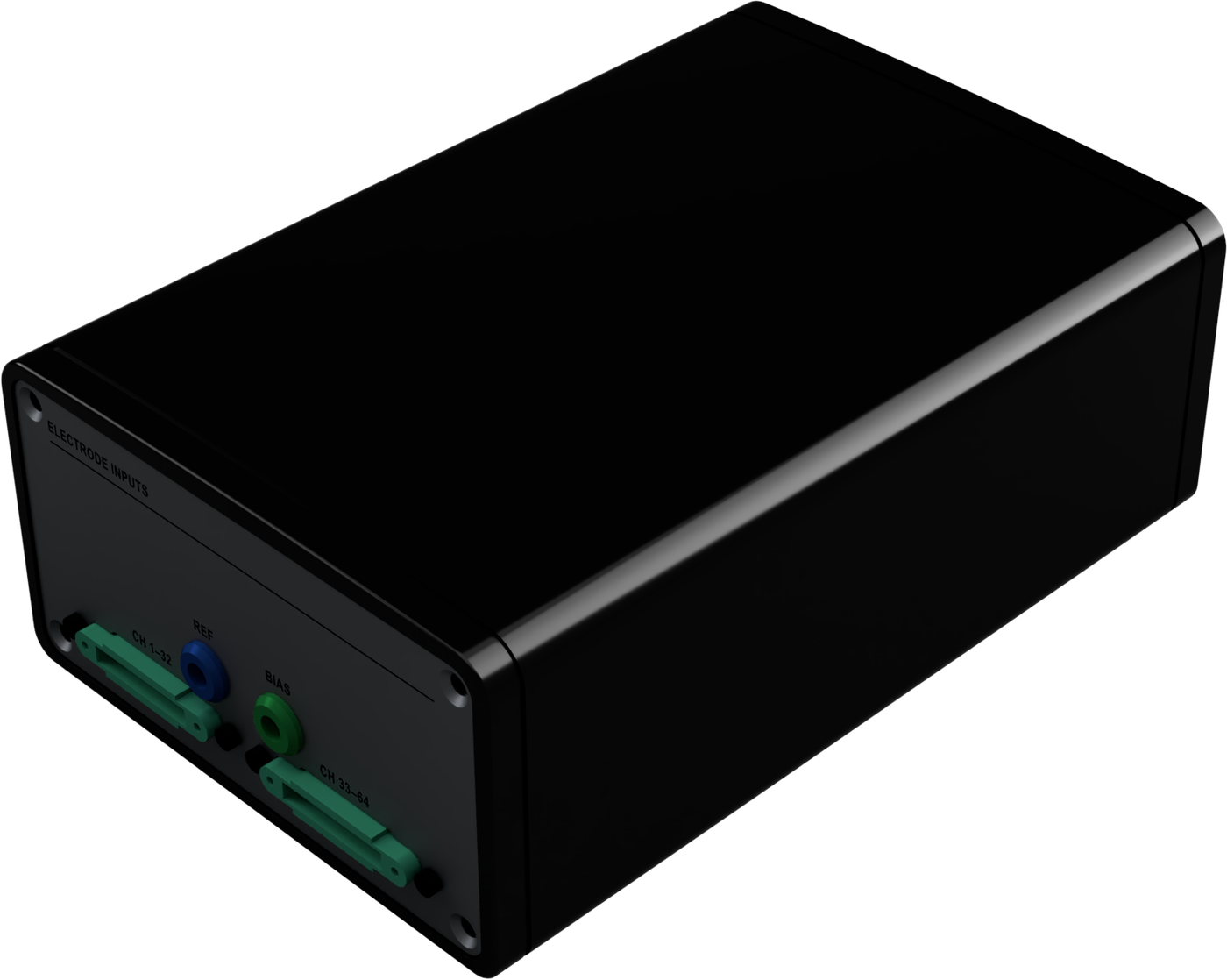
Three sensor types for one recording unit
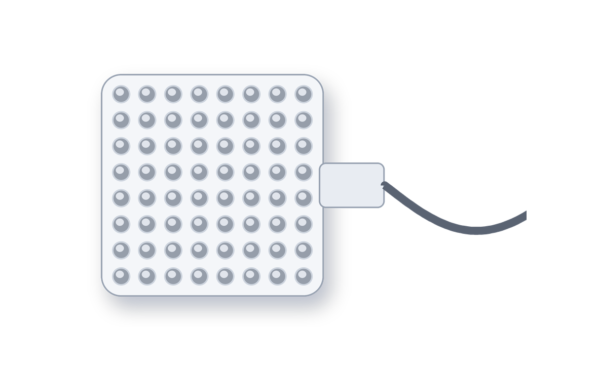
Sensor grid
A flexible patch with 64 sensors in an 8 × 8 grid. It sits on the skin over one trunk or limb muscle and maps that muscle in detail.
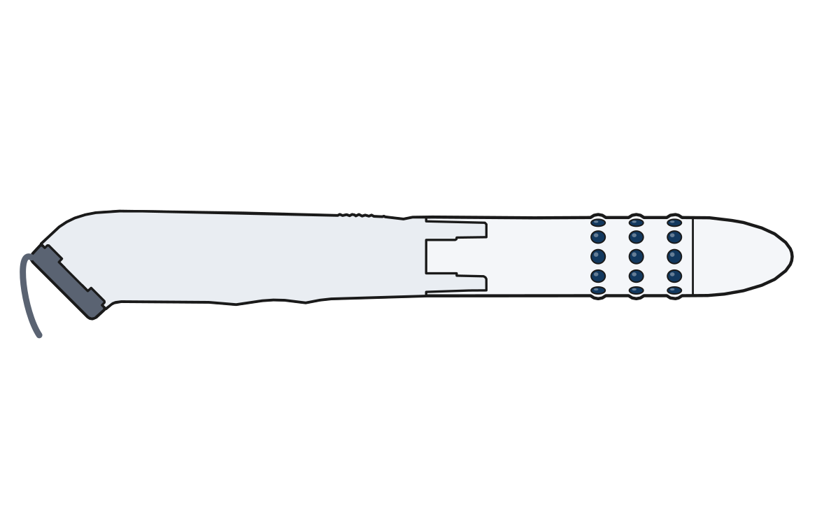
Pelvic floor probe
A single-use probe with 36 sensors in 3 rings around it. It is placed in the vagina or rectum and records the pelvic floor muscles and the anal sphincter.
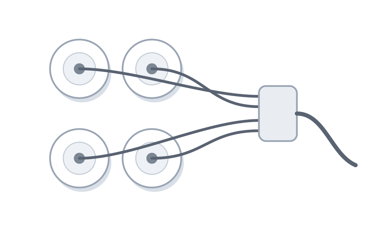
Skin sensors
Stick-on sensors in pairs, one pair per muscle. With the probe, up to 28 pairs can record the hip, trunk and leg muscles at the same time.
Maps show where muscles are active
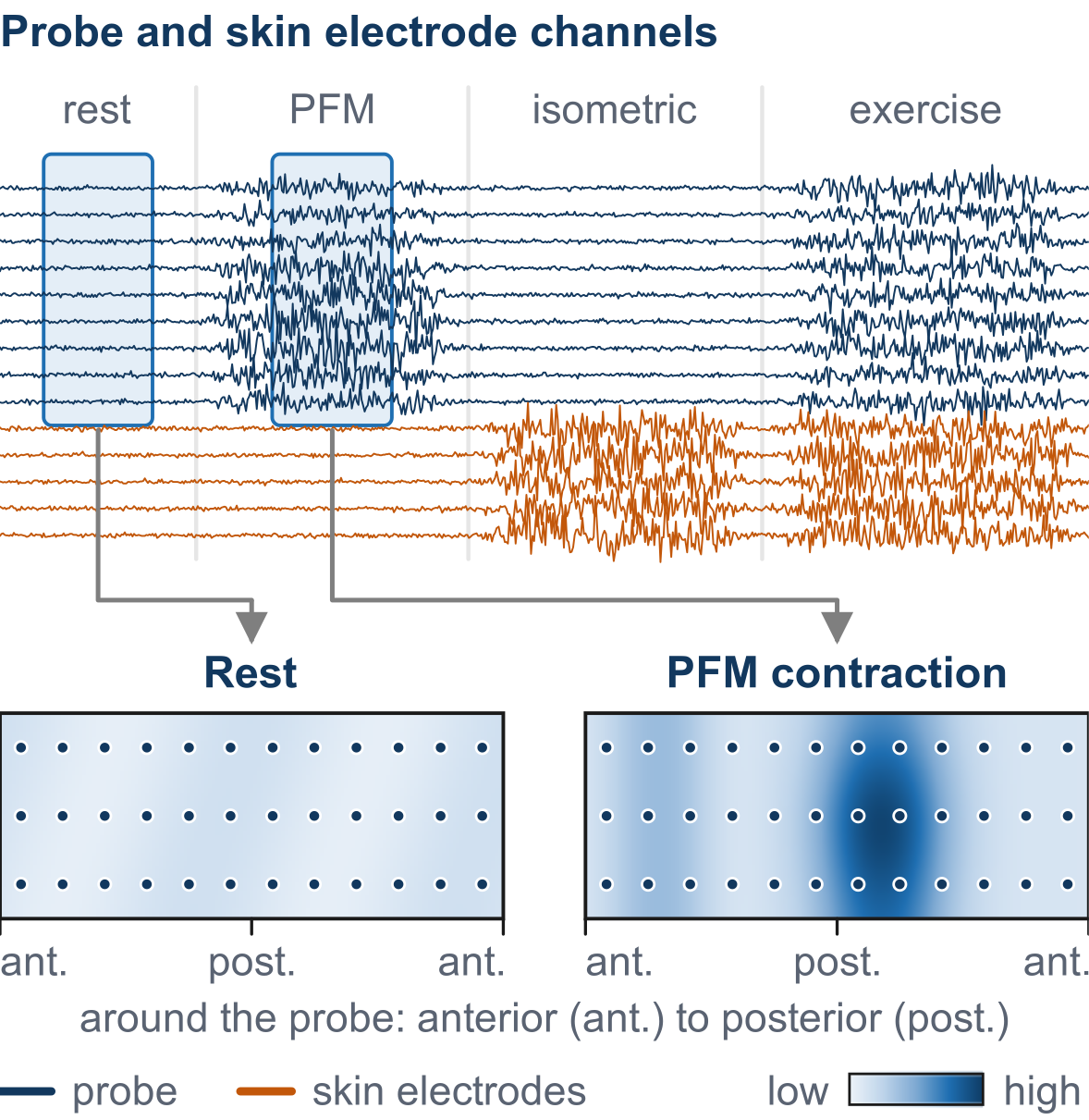
During a session, the person rests, squeezes the pelvic floor, holds steady contractions and does rehabilitation exercises while all channels record together.
For each moment, the software maps the activity around the probe. The map shows which part of the pelvic floor is working, from front to back. The skin sensors show which hip, trunk and leg muscles are active at the same time.
Single motor units from the skin
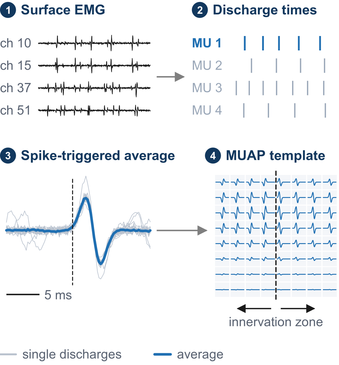
A motor unit is one nerve cell and the muscle fibers it controls. Each sensor picks up many motor units at once. The software separates them and finds when each one fires. Averaging the signal at those moments gives the shape of each motor unit across the grid.
This shows how often each motor unit fires, how large its signal is, and how fast the signal travels. It also estimates how many motor units still work in a muscle. In a 2023 study by Liu and colleagues, stroke survivors had fewer working motor units in the stiff biceps than on the other side.
Where nerves join a muscle
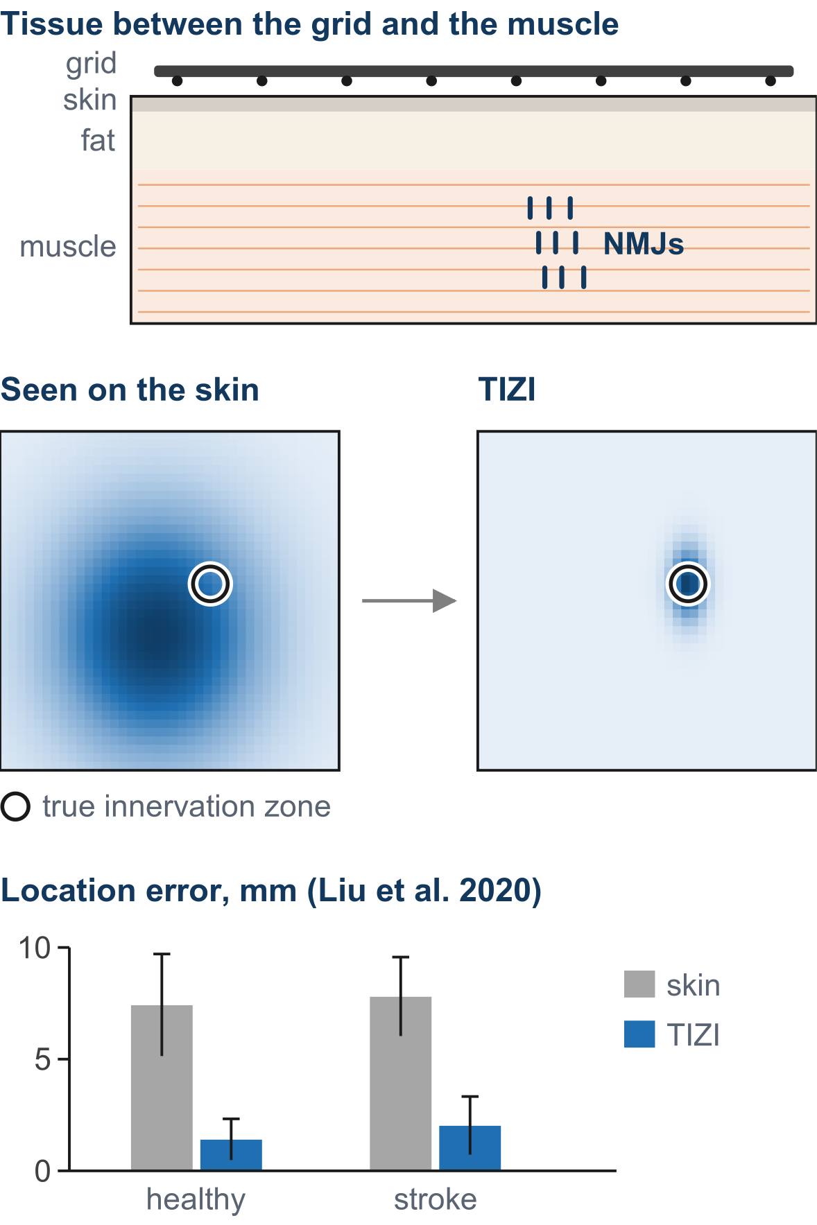
Nerves join each muscle in a narrow band. Skin and fat between the muscle and the sensors can shift where this band appears. The software is being designed to correct for these layers.
Botulinum toxin is a medicine injected to relax stiff muscles after a stroke. It acts where nerves join the muscle. Researchers are studying whether maps of this band can help plan injections. In a 2020 study by Liu and colleagues, the band was located within about 2 mm, compared with about 8 mm when judged from the skin alone.
How muscles work together
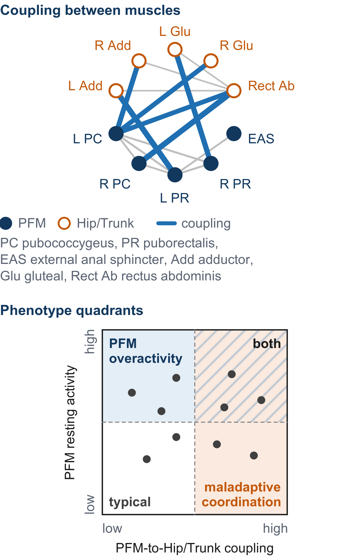
The software measures how closely each pair of muscles works together and groups them into networks. HillMed holds a patent on this method.
In a 2021 study by Houston and colleagues of 8 shoulder and elbow muscles, healthy adults had 4 networks. Stroke survivors had 3 or 2, with fewer networks as stroke effects increased.
For the pelvic floor, the software compares resting pelvic floor activity and how strongly the pelvic floor works with hip and trunk muscles against ranges from people without symptoms. It places each recording in one of four groups: typical, high resting activity, pelvic floor working with hip and trunk muscles, or both.
At a glance
| Channels | 64, recorded at the same time |
| Sensor grid | 8 × 8 sensors |
| Pelvic floor probe | 36 sensors in 3 rings, about 1 inch (25.6 mm) wide, single use, vaginal or rectal |
| Skin sensors | Up to 28 pairs together with the probe |
| Software | Activity maps, single motor units, where nerves join a muscle, muscle networks, four-group profile |
| Research areas | Urinary incontinence, pelvic organ prolapse, pelvic pain, stroke, spasticity, rehabilitation |
| Status | In development. Not for sale. Not cleared or approved by the FDA or any other regulator. |
Stay in touch
Researchers can join our interest list to hear when a demonstration is ready.