Unveiling the Language of Your Muscles
Our bodies are intricate networks of nerves and muscles which must communicate with each other to make our bodies move. This communication relies on tiny electrical signals that orchestrate the complex dance of muscle contraction. Understanding this electrical language is key to knowing how our muscles function when healthy and behave when affected by disease or injury.
Nerves and Muscles Working Together
The journey of movement begins in the brain, which sends commands in the form of electrical impulses. These impulses travel down the spinal cord and then branch out through specialized nerve cells called motor neurons. Each motor neuron acts like a dedicated messenger, carrying its signal to a specific group of muscle fibers.
When the electrical signal from the motor neuron reaches these muscle fibers, it triggers them to contract, or shorten, generating force and producing movement. This entire process is a continuous flow of electrical information, a vital dialogue that keeps our bodies in motion.
The Motor Unit
To understand muscle function at a more granular level, we need to understand the “motor unit” (MU). An MU consists of a single motor neuron and all the individual muscle fibers it connects to, or “innervates.“. Think of it as the smallest functional team within the muscle that the brain can independently control.
When a motor neuron fires its electrical signal, all the muscle fibers within its unit contract together, almost simultaneously. This coordinated electrical activity of the muscle fibers within an MU generates a distinct electrical signature known as a Motor Unit Action Potential (MUAP).
Each time an MU fires, it produces a MUAP. A series of these firings, or MUAPs, from a single motor unit over time forms a pattern, often referred to as an Innervation Pulse Train (IPT). This IPT is like a coded message which tells us the timing and frequency of the MU’s contribution to the muscle’s overall effort.
What is Electromyography?
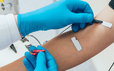
Electromyography, or EMG, is a technique used to listen in on this electrical conversation within our muscles. By placing sensors, called electrodes, near or within a muscle, clinicians and researchers can record the electrical signals produced when muscles are active.
Traditionally, EMG could involve inserting fine needle electrodes directly into the muscle (intramuscular EMG) to get very localized information, or placing a few electrodes on the surface of the skin (surface EMG) to get a more general idea of the muscle’s activity.
Moving towards High-Density Surface EMG
Surface EMG (sEMG) offers a non-invasive way to measure muscle activity by simply placing electrodes on the skin over a muscle. High-Density sEMG (HD sEMG) takes this concept a significant step further. Instead of using just one or a few electrodes, HD sEMG employs often dozens of small electrodes arranged in a closely packed grid or array that is placed on the skin over the muscle.
Imagine trying to understand the conversations in a crowded room. Traditional sEMG with a few electrodes is like having one or two microphones; you might hear the overall hum of voices, but it’s difficult to distinguish individual speakers.
HD sEMG, on the other hand, is like having an array of many microphones strategically placed throughout the room. Each microphone picks up a slightly different version of the sound, and by analyzing the signals from all these microphones, it becomes possible to start isolating individual conversations.
Similarly, the dense array of electrodes in HD sEMG captures a much richer and more detailed map of the electrical activity across the muscle surface.
This shift to HD sEMG is also about capturing the spatial distribution of the MUAPs as they propagate under the skin. Different MUs, due to their varying anatomical locations (depth and position within the muscle), will generate electrical fields that project differently onto the overlying grid of electrodes. This spatial richness, the unique electrical fingerprint each MU leaves on the electrode array, is a critical piece of information.
Without this detailed spatial sampling, distinguishing the signals from MUs that are firing closely in time or are located near each other would be significantly more challenging, much like trying to pinpoint the source of a sound with only one ear.
Furthermore, the non-invasive nature of HD sEMG represents a profound advantage. Unlike needle EMG, which can be painful and is limited in how much of the muscle it can sample, HD sEMG is comfortable and can be applied repeatedly without discomfort. This makes it exceptionally well-suited for studies involving children or other vulnerable populations, for long-term monitoring scenarios and for applications like biofeedback or controlling prosthetic devices, where patient comfort and ease of use are paramount for practical success.
The following table provides a comparison to illustrate the progression in what we can learn from muscle signals:
Table 1: Understanding Muscle Signals: Traditional EMG vs. HD sEMG (Leading to Decomposition)
| Feature | Traditional Surface EMG | High-Density Surface EMG (Raw) | HD sEMG (Decomposed) |
|---|---|---|---|
| Number of Electrodes | Few (e.g., 1–4) | Many (e.g., 32–128) | Many (e.g., 32–128) |
| Type of Information | Global muscle activity, overall amplitude/timing | Detailed map of electrical activity over muscle surface | Individual motor unit firing times (IPTs) and shapes (MUAPs) |
| Level of Detail | Low, “blurry” view | Moderate, potential for detail but signals still mixed | High, “clear” view of individual units |
| Primary Insight | General muscle activation, overall fatigue assessment | Spatial distribution of activity, areas of high/low activity | Neural drive to muscle, specific MU recruitment strategies, coordination between MUs |
| Analogy | Listening to a stadium with one microphone | Having multiple microphones in a stadium, hearing distinct sections | Isolating individual voices from the stadium crowd |
This table shows that while raw HD sEMG provides more data, the real breakthrough comes from the subsequent step: decomposition.
What is HD sEMG Decomposition?
When you use a muscle, it’s not just one motor unit working in isolation. Dozens, sometimes hundreds, of MUs can be active simultaneously, each firing at its own rate and intensity to contribute to the overall force and precision of the movement. The electrical signals from all these concurrently active MUs blend together.
The “Unmixing” Process: What is Decomposition?
HD sEMG decomposition is a sophisticated set of computational techniques, often involving advanced signal processing and algorithms, designed to tackle this “unmixing” problem. Its primary goal is to take the complex, mixed HD sEMG signal recorded from the skin surface and identify and separate the individual “voices” within it. These voices are the constituent MUAPs and their corresponding firing times (the IPTs) for as many distinct MUs as possible.
This process is often referred to as blind source separation. The process is blind because, in most cases, the system doesn’t have prior knowledge of exactly what each MU’s individual signal looks like, nor does it know precisely when each MU is going to fire. The algorithms must intelligently deduce this information from the patterns and consistencies present in the mixed, multi-channel recordings.
The success of this blind separation hinges on the fact that MUAPs from different MUs, when viewed across the high-density electrode array, tend to have sufficiently distinct spatiotemporal characteristics, subtle differences in their shape or how their electrical activity spreads across the skin surface.
These distinctions allow the algorithms to differentiate them, even when their activity overlaps in time. Hence, the density of electrodes is crucial, as it provides multiple, slightly different “views” of each MU’s activity, enhancing the ability of algorithms to tease them apart.
What We Learn from Decomposed Signals
Once the HD sEMG signal has been successfully decomposed, we gain access to the unique “signature” of each identified active motor unit. This signature typically includes two key pieces of information:
- Its Firing Pattern (Innervation Pulse Train – IPT): This tells us precisely when, and how frequently, each MU discharged. This sequence of firing times is a direct reflection of the commands being sent from the motor neuron in the spinal cord to that specific MU.
- Its Waveform (Motor Unit Action Potential – MUAP): This is the characteristic electrical shape of that MU’s signal as detected by the array of electrodes. The shape, amplitude and duration of the MUAP can provide valuable clues about the physiological properties of the MU, such as its relative size (number of muscle fibers it contains), the health of its muscle fibers and its location relative to the electrodes.
The Crucial Role of Signal Clarity
The ability to accurately decompose HD sEMG signals is heavily dependent on the quality of the initial recordings. Electrical “noise” can contaminate the delicate MUAP signals and stem from unwanted signals from various sources including:
- Other nearby electrical devices
- Movement of the electrodes on the skin
- Instability at the electrode-skin interface
This noise makes it harder to hear the signal clearly. If too strong, it can obscure the MUAPs, making it difficult or impossible for decomposition algorithms to identify them correctly.
Why Does Decomposing Muscle Signals Matter?
The ability to non-invasively peer into the detailed workings of individual motor units through HD sEMG decomposition opens up a wealth of possibilities for understanding human movement.
A Non-Invasive Window into the Nervous System’s Control
Perhaps the most profound significance of HD sEMG decomposition is that it provides a direct, non-invasive window into the neural control of muscles. For the first time, we can observe, in conscious humans performing natural movements, the precise output of individual spinal motor neurons.
This allows researchers and clinicians to study how the brain and spinal cord orchestrate muscle activity to produce force, control movement and learn new skills, all without the need for invasive procedures like needle insertions into muscles or nerves. This capability offers direct insights into the strategies the nervous system employs, revealing how it recruits different MUs, modulates their firing rates and coordinates their activity to meet the demands of various tasks.
Fueling Discoveries in Movement Science and Physiology
For researchers in fields like kinesiology, biomechanics and motor control, HD sEMG decomposition is an invaluable tool for exploring the fundamental principles of how our bodies move. It can be used to investigate:
- Motor Learning: How does the nervous system change its MU control strategies as we learn a new motor skill?
- Muscle Fatigue: What happens at the individual MU level when a muscle fatigues? Do firing rates decline, does MU recruitment change, or do MUAP shapes alter?
- Adaptation: How do MUs adapt to strength training, endurance exercise, periods of disuse (like bed rest), or the natural process of aging?
- Coordination: How does the nervous system coordinate the activity of MUs within a single muscle, or across multiple synergistic muscles, to produce smooth and efficient movements?
Transforming Clinical Understanding of Neuromuscular Conditions
In the clinical realm, HD sEMG decomposition holds immense promise for improving the diagnosis, monitoring and understanding of a wide range of neuromuscular conditions. These include:
- Neurodegenerative diseases: Such as amyotrophic lateral sclerosis (ALS), where motor neurons progressively degenerate.
- Peripheral neuropathies: Conditions affecting the peripheral nerves, for example, due to diabetes or injury.
- Myopathies: Diseases primarily affecting the muscle fibers themselves, like muscular dystrophies.
- Spinal cord injuries: Where communication between the brain and muscles is disrupted.
- Movement disorders: Such as Parkinson’s disease or tremors.
By examining the behavior of individual MUs – their number, firing rates, recruitment thresholds and MUAP characteristics – clinicians may be able to detect subtle abnormalities much earlier than is possible with traditional clinical examinations or global EMG measures.
For instance, the ability of advanced systems to identify a higher number of unique MUs, particularly at low force contraction levels (e.g., ≤20% of maximum voluntary contraction), suggests a heightened sensitivity to the nuances of muscle activation that might be crucial for early diagnosis or for tracking subtle changes. This is particularly important because many activities of daily living involve fine motor control at these lower force levels, and the initial signs of some neuromuscular diseases might first appear as alterations in the behavior of these low-threshold MUs.
The capacity to non-invasively track the characteristics of individual motor units over extended periods – for example, throughout the progression of a disease or during a rehabilitation program – represents a significant leap forward. Previously, assessing such longitudinal changes at the MU level often necessitated repeated invasive procedures, which are impractical for routine monitoring and can be poorly tolerated by patients. HD sEMG decomposition offers a patient-friendly alternative that could revolutionize how we monitor disease and evaluate the effectiveness of treatments.
Furthermore, the rich, multi-dimensional dataset generated by decomposed HD sEMG – encompassing the firing patterns and MUAP characteristics of numerous MUs – is ideally suited for analysis with advanced computational tools, including machine learning and artificial intelligence. These approaches could potentially uncover complex biomarkers or subtle patterns of MU dysfunction that are indicative of specific diseases, stages of disease, or responses to therapy, patterns that might be entirely missed by simpler, aggregate EMG measures or visual inspection alone. This opens avenues for more objective and data-driven clinical decision-making.
How Can HD sEMG Decomposition Be Used?
The detailed insights provided by HD sEMG decomposition are paving the way for a host of innovative applications across medicine, rehabilitation and human-machine interaction. Let’s unpack some of the big ones:
- Sharpening Diagnosis: Traditional diagnostic approaches might identify muscle weakness or abnormal global EMG patterns. However, HD sEMG decomposition can delve deeper to help pinpoint the underlying cause of such issues. For example, if a patient presents with weakness, decomposition could reveal whether this is due to a reduced number of MUs being successfully activated by the nervous system, whether the active MUs are firing at inappropriately slow rates, or if the MUAPs themselves have abnormal shapes (e.g., prolonged duration or reduced amplitude), which might suggest problems with the muscle fibers or their innervation. This detailed information can be crucial in distinguishing between conditions that primarily affect the nerves (neuropathies) and those that primarily affect the muscles (myopathies), guiding further diagnostic tests and treatment strategies.
- Tracking Disease Progression: The ability to non-invasively quantify changes in individual MU function over time offers a powerful tool for objectively monitoring disease progression or the effects of an intervention. Instead of relying solely on subjective patient reports or gross measures of strength, clinicians could track specific MU parameters. For instance, in a patient recovering from a nerve injury, the re-appearance of MUAPs, increases in their firing rates, or normalization of their shapes could serve as objective markers of reinnervation and functional recovery. Conversely, in a progressive neurodegenerative disease, a decline in the number of active MUs or changes in their firing characteristics could provide early warnings of worsening pathology.
- Informing More Targeted Therapies: Neuromuscular diseases often do not affect all MUs within a muscle uniformly. Some conditions might preferentially target smaller, low-threshold MUs, while others might impact larger, high-threshold units, or affect MUs based on their fiber type (e.g., fast-twitch vs. slow-twitch). HD sEMG decomposition can help identify such selective vulnerabilities. This nuanced understanding of which specific MU populations are dysfunctional is a critical first step towards developing more targeted therapies. If clinicians can identify the precise nature and location of MU deficits, interventions can, in principle, be designed to address those specific issues, moving beyond generic muscle strengthening or symptomatic relief.
- Personalized Biofeedback and Rehabilitation: One of the most exciting applications is in the realm of real-time biofeedback. Imagine a patient who has difficulty activating certain muscles after a stroke or injury. With HD sEMG decomposition, it’s possible to provide them with direct, real-time visual or auditory feedback about the activity of specific MUs they are trying to control. They could, for instance, play a simple computer game where success depends on activating and modulating the firing of a target MU that was previously underactive. This type of highly specific biofeedback can facilitate motor learning and help patients regain control over weakened or uncoordinated muscles far more effectively than feedback based on global muscle effort alone.
- Objectively Measuring the True Impact of Therapies: When evaluating new drugs or rehabilitation protocols, HD sEMG decomposition can provide objective, physiological measures of treatment efficacy that go beyond simple strength tests or functional scores. Researchers can assess whether a therapy leads to an increase in the number of recruited MUs, enhances their firing rates, improves the synchrony of their discharges (if desired), or restores more normal MUAP characteristics. This detailed information can help to understand how a therapy is working at the neuromuscular level and optimize treatment protocols.
- Facilitating More Intuitive and Dexterous Prosthetic Limbs: For individuals with limb loss, HD sEMG decomposition offers the potential to create prosthetic devices that are controlled with unprecedented naturalness and dexterity. By decoding the intended movement commands from the firing patterns of many individual MUs in the residual muscles of the stump, it’s possible to extract a much richer and more detailed control signal than what can be obtained from traditional EMG. This high-bandwidth information can then be used to command multi-degree-of-freedom prosthetic hands, wrists and elbows, allowing users to perform complex grasping and manipulation tasks more intuitively. The ability to simultaneously decode signals from multiple MUs is key here; different combinations and firing rates of these MUs can be mapped to a wide array of prosthetic functions, offering a level of control that approaches that of a natural limb.
- Guiding Advanced Assistive Devices for Paralysis: Similarly, for individuals with paralysis due to conditions like spinal cord injury or stroke, but who retain some residual voluntary control over a few MUs, decomposed HD sEMG can translate these faint or isolated muscle signals into reliable commands for controlling external devices. This could include powered wheelchairs, environmental control systems, communication aids or even robotic exoskeletons designed to assist with movement. The non-invasive nature of HD sEMG makes it an attractive option for long-term human-machine interfacing.
- Optimizing Athlete Performance: In sports science and athletic training, HD sEMG decomposition can provide detailed insights into muscle recruitment strategies during specific movements. Coaches and athletes could use this information to identify inefficient muscle activation patterns, optimize training regimens to enhance performance, or detect early signs of muscle imbalances or fatigue patterns that might predispose an athlete to injury.
- Enhancing Ergonomics and Workplace Safety: In occupational health, HD sEMG can be used to study muscle activation and loading during various work tasks. By understanding how different MUs are utilized, and which ones are prone to overuse or fatigue, ergonomists can help design tools, workstations and work processes that minimize the risk of musculoskeletal disorders and repetitive strain injuries.
For many of these real-world applications, particularly those involving continuous control like prosthetics or dynamic activities like sports, the robustness of the HD sEMG system and its decomposition algorithms is critical. The system must perform reliably despite challenges like sweat affecting electrode contact, movement artifacts or changing electrical interference from the environment.
Advances in developing ultra-low-noise amplifier hardware and robust decomposition algorithms that can function effectively even when the recording environment isn’t perfect are therefore essential enablers for translating this technology from the laboratory into everyday use.