Listening to Our Muscles
At its core, electromyography (EMG) is a way of listening to the electrical signals produced by our muscles when they contract or are at rest. When the brain tells a muscle to work, electrical impulses travel along nerves to the muscle fibers, causing them to activate.
EMG techniques use electrodes, which are small sensors, to detect these electrical signals, known as electromyograms. By analyzing these signals, we can gain valuable insights into muscle health, how muscles are controlled by the nervous system, and how they respond to various conditions or treatments.
There are two techniques used today to study muscle function: conventional surface electromyography (sEMG) and high-density surface electromyography (HD-sEMG). For the uninitiated, it can be easy to assume that HD-sEMG is simply a higher quality reading of our muscle’s signals. However, that assumption would be misguided.
Let’s dig into the definitions and differences between both techniques, including some practical and meaningful instances where each can be applied:
Conventional Surface Electromyography (sEMG)
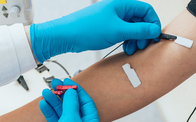
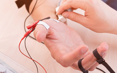
Conventional surface EMG (sEMG) is a widely used, non-invasive method to assess muscle activity. It involves placing electrodes on the surface of the skin over the muscle or muscles of interest.
This technique captures the overall electrical activity generated by the muscles underneath the electrodes. It provides a global measure of when a muscle is active, the general intensity of its contraction—and sometimes how different muscles coordinate during movements. For example, sEMG has been used to study intermuscular coherence (IMC) to understand how different muscles in the upper arm work together, or don’t, in individuals who have had a stroke.
Typical Electrode Setup
In conventional sEMG, a relatively small number of electrodes are used for each muscle being studied. Often, this involves a bipolar configuration, where two detection electrodes are placed along the length of the muscle, typically over the muscle belly, and a reference electrode is placed on an electrically neutral area (like a bony prominence). The specific placement aims to be between a motor point (where the nerve enters the muscle) and the tendon, avoiding these specific areas to get a clearer signal from the bulk of the muscle fibers. The number of channels (pairs of electrodes) corresponds to the number of muscles or muscle sites being monitored.
How Does it Work?
When muscle fibers activate, they generate electrical potentials. Conventional sEMG electrodes detect the sum of these electrical potentials from many muscle fibers in the vicinity of the electrodes. The recorded signal represents the overall electrical output of a significant portion of the muscle. This signal is then typically amplified and filtered to remove noise and highlight the muscle’s activity pattern.
What Information Can We Get from Conventional sEMG?
Therapists and their patients can gain multiple, helpful insights from traditional sEMG. These include:
- Muscle Activation Timing: When a muscle turns on and off during a movement or task.Amplitude/Intensity of Contraction: A general idea of how strongly a muscle is contracting (though this is influenced by many factors).
- Muscle Fatigue: Changes in the EMG signal characteristics (like frequency content) can indicate muscle fatigue.
- Inter-Muscular Coordination: By recording from multiple muscles simultaneously, sEMG can be used to study how different muscles work together, for instance, by analyzing intermuscular coherence, which reflects common neural input to different muscles.
Analogy: The Orchestra with Few Microphones:
Think of a muscle as an orchestra with many musicians (muscle fibers). Conventional sEMG is like placing a few microphones in the concert hall. You can tell when the orchestra starts and stops playing, get a sense of how loud the music is overall, and perhaps identify if the brass section is playing with the strings.
However, you wouldn’t be able to hear each individual instrument clearly or pinpoint exactly where a specific sound is coming from within the orchestra.
Introducing High-Density Surface Electromyography (HD-sEMG)
High-Density Surface Electromyography (HD-sEMG) is a more advanced technique that provides a much more detailed and spatially resolved view of muscle activity compared to conventional sEMG.
HD-sEMG utilizes a significantly larger number of electrodes, arranged in a closely packed grid or array, placed over the skin surface of a muscle or muscle group. This high density of sensors allows for the capture of “abundant spatiotemporal PFM activity information,” meaning it records muscle activity across both space (the area covered by the grid) and time with high fidelity.
Typical Electrode Setup
HD-sEMG systems employ grids or arrays containing many small electrodes. For example, studies have used 64-channel (8×8) grids for assessing pelvic floor muscles via intravaginal or intrarectal probes , or two 8×8 grids (128 channels total) placed adjacently over the biceps brachii muscle. The inter-electrode distance (the spacing between the centers of adjacent electrodes) is typically small, for instance, 8.5 mm, 8.8 mm, or 5.7 mm, depending on the specific probe design and the direction of measurement. This close spacing is critical for its enhanced capabilities.
How Does it Work?
Each individual electrode in the HD-sEMG grid records the electrical activity from a relatively small and specific area of the muscle directly beneath it. Because there are many electrodes packed closely together, HD-sEMG effectively creates a detailed “electrical map” of muscle activity across the entire area covered by the grid. This rich dataset, often comprising signals from 64 or 128 channels, is then subjected to sophisticated signal processing techniques, such as “HD-SEMG decomposition” or “3DIZI calculation,” to extract meaningful physiological information.
What Information Can We Get from HD-sEMG?
The dense sampling of muscle electrical activity enables HD-sEMG to provide several types of information that are generally not obtainable with conventional sEMG. These include:
- Detailed Spatial Maps of Muscle Activity: HD-sEMG can generate visual representations “heat maps,” that show precisely where a muscle is most or least active, or identify specific regions of overactivity (hypertonicity). For instance, a “hypertonicity index” can be calculated, and “hypertonic zones” or “RMS mapping hotspots” can be visualized. These maps can illustrate how activity is distributed across the muscle during various tasks or even at rest.
- Identification of Innervation Zones (IZs): Innervation zones are the specific regions where motor nerves form synapses with muscle fibers, and where neuromuscular junctions are densely distributed. HD-sEMG can pinpoint these IZs by observing how electrical signals, known as Motor Unit Action Potentials (MUAPs), originate and propagate along muscle fibers. This is often done by looking for a “phase reversal” in the signals recorded by adjacent electrodes. This capability allows for the creation of “patient-specific IZ mappings,” sometimes even in three dimensions, including information about the depth of the IZ from the skin surface. Specialized techniques like 3D Innervation Zone Imaging (3DIZI) leverage HD-sEMG for this purpose.
- Analysis of Individual Motor Units (MUs): A motor unit consists of a single motor neuron (nerve cell) and all the muscle fibers it controls. HD-sEMG signals can often be “decomposed” using advanced algorithms to identify the distinct firing patterns of individual motor units within the muscle. This decomposition allows researchers and clinicians to assess properties like MU firing rates, the way MUAPs travel along muscle fibers (propagation), the shape and size of MUAPs (morphology), and even to estimate the number of active motor units (e.g., using a Motor Unit Number Index or MUNIX).
- Assessment of Muscle Fiber Signal Propagation: The technique can track how the electrical signal (MUAP) initiated at an IZ travels along the muscle fibers in opposing directions. This information is valuable for understanding muscle fiber health and the integrity of neuromuscular transmission.
- Detection of Asymmetry and Localized Changes: Due to its high spatial resolution, HD-sEMG can detect subtle differences in muscle activity or innervation patterns between the left and right sides of the body, or within different compartments of a single muscle or muscle group. For example, it has been used to calculate an “asymmetry index” for the external anal sphincter.
The shift towards HD-sEMG signifies a substantial advancement in our ability to study muscles. The high density of electrodes allows for a transition from an averaged view of muscle function to a spatially explicit understanding. This capability to generate detailed maps of activity, pinpoint critical areas like innervation zones, and even learn about the behavior of individual motor units non-invasively is transformative. It’s akin to moving from a general understanding that a muscle is “working” or “not working” to being able to see the intricate details of how and where it’s functioning or malfunctioning.
Analogy: The Orchestra with Individual Microphones and 3D Sound Mapping
Revisiting the orchestra analogy, HD-sEMG is like placing a dedicated microphone on every single instrument. Furthermore, it’s like having a sophisticated sound engineer who can create a detailed map showing exactly which instrument is playing, how loudly, what notes they are playing, and if they are positioned correctly within the orchestra.
For deeper muscles or when 3D information is needed (as with 3DIZI), it’s as if this engineer also has “X-ray hearing” to determine the depth and precise 3D location of each sound source.
sEMG vs. HD-sEMG: Understanding the Key Distinctions
The core difference between conventional sEMG and HD-sEMG lies in the density of electrodes and, consequently, the richness and spatial detail of the information they can capture. While conventional sEMG provides a valuable global overview, HD-sEMG offers a microscopic, high-resolution map of muscle activity.
The following table provides a side-by-side comparison of key features:
| Feature | Conventional sEMG | High-Density sEMG (HD-sEMG) | Why this Matters |
|---|---|---|---|
| Typical # of Electrodes | Few (e.g., 2–4 per muscle/channel) | Many (e.g., 64, 128, or more in a grid/array) | More “listening posts” mean HD-sEMG can pick up much finer details about what different parts of the muscle are doing. |
| Electrode Density & Arrangement | Lower density, often placed broadly along the muscle belly. | High density, with electrodes closely packed in a grid or array configuration. | Closer “listening posts” allow HD-sEMG to distinguish signals from very small, nearby areas of the muscle, like having more pixels in a digital image. |
| Primary Information Captured | General muscle on/off timing, overall activity level or force estimation. Sometimes used to assess coordination between different muscles. | Detailed spatial maps of activity across the muscle, precise location of innervation zones (IZs), characteristics of individual motor unit action potentials (MUAPs) including their firing patterns and propagation, and muscle fiber conduction velocity. | Conventional sEMG gives the “big picture”: is the muscle active? HD-sEMG provides a “detailed blueprint”, how and where is the muscle active. |
| Spatial Resolution | Low to moderate (sees the forest, or a large group of trees). | High (sees individual trees, and sometimes even the leaves on the branches). | HD-sEMG can pinpoint problems or areas of interest to very specific locations within a muscle, while conventional sEMG might only indicate a general issue in the ability to activate the entire muscle. |
| Ability to Identify IZs | Generally No. The limited number of electrodes and their spacing do not typically allow for the detection of signal propagation patterns necessary to locate IZs. | Yes, often with high precision, and can even map them in three dimensions (including depth). | Knowing exactly where nerves connect to muscles (IZs) is vital for understanding nerve-related muscle problems and for guiding treatments like Botox injections to ensure they are most effective. HD-sEMG can find these critical spots. |
| Motor Unit Detail | Limited; can provide general information about overall motor unit recruitment. | Can often “decompose” the complex EMG signal to study the firing patterns and behavior of individual motor units. | HD-sEMG can effectively “listen in” on individual motor units, helping to understand how the brain is controlling the muscle at a very fine level. This is extremely useful for detecting subtle nerve damage, muscle disease, or changes during recovery. |
| Key Clinical Uses/Advantages | Biofeedback, general assessment of muscle function (e.g., in sports science), gait analysis, monitoring overall muscle activation. | Precise diagnosis of localized muscle dysfunction (e.g., specific trigger points or hypertonic areas), personalized treatment guidance (e.g., for BoNT injections, targeted physical therapy), objective mapping of muscle hypertonicity or weakness, advanced neurophysiological research into motor control and disease. | Conventional sEMG is good for general functional checks. HD-sEMG excels at identifying specific, localized issues and guiding precise treatments, making interventions more effective and personalized. It can turn subjective patient complaints into objective, visual maps that clinicians can use. |
Information Depth
Conventional sEMG is excellent for answering the question, “Is the muscle working, and roughly how much?” HD-sEMG, on the other hand, is better suited to answer more detailed questions like, “How, precisely where, and how well is this muscle working, down to specific regions, nerve connections, and even groups of muscle fibers controlled by single nerves?”.
Analogy: Diagnosing a Car Problem
If your car engine is making a strange noise, conventional sEMG is like a mechanic listening to the engine with their ear to get a general idea if it’s running rough. HD-sEMG is like plugging the car into a sophisticated diagnostic computer. This computer can tell you exactly which cylinder is misfiring, check the performance of individual sensors, map out the engine’s efficiency in different areas, and pinpoint the source of the problem with much greater accuracy.
RHD-sEMG in Real-World Examples
The detailed information provided by HD-sEMG translates into significant, tangible benefits for both patients and clinicians across a range of medical conditions. Its ability to precisely map muscle activity and locate key neurophysiological landmarks offers new avenues for diagnosis and treatment.
Example 1: Understanding and Treating Pelvic Floor Muscle Problems (e.g., Hypertonicity, Pain, Incontinence)
The Challenge: The pelvic floor muscles form a complex, multi-layered structure that is crucial for functions like bladder and bowel control, sexual function, and core stability. Assessing these deep-lying muscles accurately can be challenging. Conditions like chronic pelvic pain, pelvic floor hypertonicity (excessive muscle tension), and urinary or fecal incontinence are common, but pinpointing the exact muscular component of the problem using manual examination alone can be difficult and subjective.
HD-sEMG in Action: Specialized intravaginal or intrarectal HD-sEMG probes, equipped with 64-channel electrode arrays, can create detailed maps of pelvic floor muscle activity. These tools can:
- Objectively identify and map “hotspots” or “hypertonic zones” where muscles are excessively active at rest. This information can be correlated with patient-reported pain levels, providing an objective measure of hypertonicity. For example, a “hypertonicity index” derived from HD-sEMG has been shown to be higher in women with pelvic floor hypertonicity.
- Assess muscle weakness or impaired activation patterns in conditions like stress urinary incontinence.
- Precisely map the innervation zones (IZs) of individual pelvic floor muscles. This is critical because the location of these nerve-muscle connections can vary significantly between individuals, and targeting these IZs is important for treatments like Botulinum toxin (BoNT) injections.
- Detect functional asymmetries, for instance, in the external anal sphincter, which can occur after events like childbirth and contribute to incontinence. An “asymmetry index” derived from HD-sEMG can quantify these imbalances.
Benefit: HD-sEMG offers a more accurate and objective diagnosis of the specific muscular dysfunction within the muscle. This moves beyond subjective palpation findings. This precision allows for highly personalized treatment strategies, such as guiding physical therapists to target specific overactive or weak muscles, or enabling physicians to perform more precise injections directly into the identified IZs of hypertonic muscles. This targeted approach may improve treatment efficacy and potentially reduce side effects.
Example 2: Improving Precision in Treating Muscle Spasticity (e.g., After Stroke)
The Challenge: Muscle spasticity, characterized by muscle tightness, stiffness, and involuntary spasms, is a common and disabling consequence of conditions like stroke or cerebral palsy. BoNT injections are a cornerstone of spasticity management, working by blocking nerve signals to the affected muscles, thereby inducing relaxation.
However, the effectiveness of BoNT is highly dependent on injecting it into the optimal locations within the muscle specifically, near the innervation zones where the toxin can best exert its effect.
Traditional methods for guiding these injections, such as manual palpation, electrical stimulation to find motor points (which are not always the same as IZs), or even standard ultrasound imaging (which shows anatomy but not nerve activity), may not be precise enough to consistently locate these IZs. Inaccurate injections can lead to suboptimal outcomes or the need for higher, potentially problematic, doses.
HD-sEMG in Action: HD-sEMG can be used to create three-dimensional maps of innervation zones in spastic muscles, a technique referred to as 3D Innervation Zone Imaging (3DIZI). This involves:
- Recording HD-sEMG signals from the spastic muscles dense electrode arrays.
- Decomposing these signals to identify MUAPs and their origins (IZs).
- Using sophisticated modeling to reconstruct the 3D locations of these IZs within the muscle volume.
- This 3D map then provides precise coordinates, including depth, to guide the BoNT injection needle directly to the identified IZs.
Benefit: Clinical studies have demonstrated that guiding BoNT injections using HD-sEMG-based 3DIZI leads to significantly better outcomes in spasticity reduction compared to standard ultrasound-guided injections.
For example, patients receiving 3DIZI-guided injections showed a greater decrease in the compound muscle action potential (CMAP) amplitude (a measure of overall muscle electrical excitability) and a larger reduction in muscle activation volume (MAV). Specifically, one study reported approximately a 14% greater CMAP decrease and a 27% greater MAV reduction with 3DIZI guidance. This improved precision means that the treatment is more effective, potentially allowing for the use of lower BoNT doses (which can reduce the risk of side effects and lower treatment costs) and leading to more consistent and predictable results across different patients.
Other Potential Uses:
Additional areas where HD-sEMG can offer value to various patients include:
- Rehabilitation Guidance: Providing detailed, real-time visual feedback on how specific muscles or parts of muscles are being activated during therapeutic exercises can enhance motor learning and recovery.
- Nerve Injury and Reinnervation Assessment: By tracking changes in MUAP characteristics, number (MUNIX), and morphology over time, HD-sEMG can objectively assess the extent of nerve damage and monitor the process of reinnervation (nerve regrowth and reconnection with muscle fibers) following injury or surgery.
- Sports Science and Performance Optimization: Analyzing detailed muscle activation patterns can help optimize training techniques and prevent injuries in athletes.
- Research into Neuromuscular Diseases: HD-sEMG provides a powerful, non-invasive tool for researchers to better understand the underlying pathophysiology of various neuromuscular disorders by examining alterations in motor unit behavior and muscle activation strategies. For instance, understanding complex interactions like the “PFM-Hip-Trunk muscle network” through HD-sEMG and conventional EMG could revolutionize how conditions like chronic pelvic pain are approached, moving beyond treating isolated muscles to addressing broader patterns of neuromuscular discoordination.